The science behind MINFIRE: caffeine-free nootropic
The science behind MINFIRE: caffeine-free nootropic
In this article you will read:
Share
Categories
Trending posts
The science behind MINFIRE
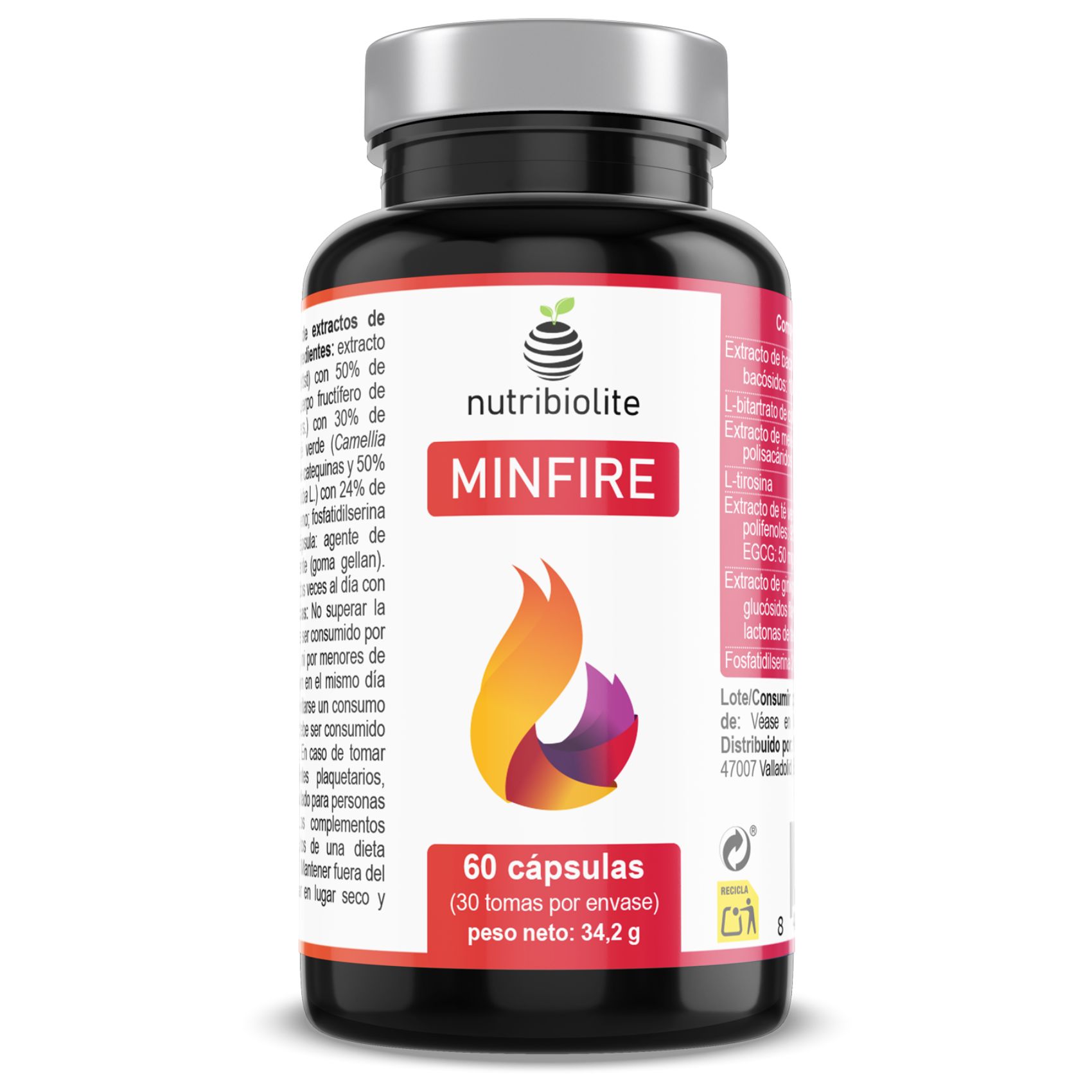
MINFIRE is a caffeine-free nootropic with extracts of selected medicinal plants, choline bitartrate, tyrosine and phosphatidylserine. It was developed to provide nutrients that participate in the cholinergic system and act in the protection of nerve cells against oxidative damage, supporting the healthy functioning of cognitive functions.
Why a caffeine-free nootropic? Many people avoid the use of nootropic supplements, due to the frequent presence of caffeine in their composition. Caffeine is the best known and most consumed stimulant on the face of the Earth, however, for many people, this substance is synonym to side effects, such as tremors, muscle spasms, anxiety, insomnia and stomach ache. Even for people less sensitive to caffeine, it is very important to limit its consumption, since caffeine is already part of our diet through the consumption of coffee or energy drinks.
The alternative: MINFIRE consists of a cocktail of adaptogens, such as the concentrated extracts of the lion’s mane mushroom (Hericium erinaceus) and the plants Bacopa monnieri, Ginkgo biloba and green tea (Camellia sinensis). In addition, MINFIRE has choline, tyrosine and phosphatidylserine in its composition, important nutrients for the health of the brain and nerve cells.
Herein, is presented a short summary of recent scientific articles published in peer-reviewed journals, that describe the properties of each one of the ingredients present in MINFIRE.
MINFIRE's unbeatable formula:
Bacopa (Bacopa monnieri L.)
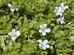
The effect of Bacoba (Bacopa monnieri L.) on memory and cognition has been extensively studied, and many excellent review articles describe its nootropic functions [1-4]. The activity has been attributed to the saponin mixture consisting of bacosides A, B and other saponins [5]. The major chemical entity shown to be responsible for the memory-facilitating action of Bacopa, are the bacoside A. It has been suggested that Bacopa, like Ginkgo biloba L., exhibits neuroprotective and cognitive enhancing effects, in part due to its capacity to modulate the cholinergic system and to contrast oxidative stress in the brain [6-9]. The brain is especially susceptible to oxidative stress because it is metabolically active, possesses high levels of pro-oxidant iron, and is composed of unsaturated lipids (prone to lipid peroxidation) [10].
The cholinergic system is composed of organized nerve cells that use the neurotransmitter acetylcholine in the transduction of action potentials. These nerve cells are activated by or contain and release acetylcholine during the propagation of a nerve impulse. The cholinergic system has been associated with a number of cognitive functions, including memory, selective attention, and emotional processing [11].
Other important property of Bacopa is its antiinflammatory activity in the brain. Neuroinflammation is thought to play a role in many central nervous system disorders including neurodegenerative diseases such as Alzheimer’s disease, and psychiatric diseases such as anxiety, depression, bipolar disorder, and schizophrenia. Long term neuroinflammation is detrimental and can lead to neurodegeneration, as seen in diseases such as Alzheimer’s disease, Parkinson’s disease, and Multiple sclerosis [12]. Bacopa has been shown in animal models to inhibit the release of the pro-inflammatory cytokines tumor necrosis factor alpha (TNFα) and Interleukin 6 (IL-6) [13]. Moreover, it was observed that alkaloid extracts of Bacopa, as well as bacoside A significantly inhibited the release of TNFα and IL-6 from activated N9 microglial cells in vitro and inhibits enzymes associated with inflammation in the brain [12].
The extract of Bacopa monnieri of MINFIRE is obtained using the last technology in plant extraction, what allows obtaining a highly pure extract and with a high concentration in bacosides. To produce our extract, a whopping 30 kilograms of Bacopa needs to be used to produce just a single kilogram of the extract. As a result, our Bacopa extract is standardized to 50% in bacosides, higher than the usually founded in other food supplements (25%-45%). Our extract meets all the quality requirements in food supplements industry, and the highly demanding criteria of Nutribiolite products.
Hericium or Lion’s mane (Hericium erinaceus Bull. Pers.)
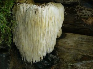
Hericium erinaceus, also known as Lion’s mane, is a very unique mushroom that is widely distributed though North America and Asia. Lion’s mane, as the name might suggest, looks like the mane of a lion. Lion’s mane has a long history as a medicine [14] and has been found to promote positive nerve and brain health. It has great potential in treating neurological disorders as it contains neurotrophic bioactive compounds that can pass through the blood–brain barrier [15, 16].
The mushroom is abundant in bioactive compounds including β-glucan polysaccharides and hericenones and erinacine terpenoides:
β-glucan polysaccharides: The β-glucan polysaccharides have been of particular interest because of the bioactivities that have been attributed to them. For example, extracellular and intracellular β-glucan polysaccharides showed a protective effect on oxidative hepatotoxicity in mice [17]. Neuroprotective effects of β-glucan polysaccharides were observed in an in vitro model of cells that were toxic from amyloid β plaque formation. β-glucan polysaccharides also promoted cell viability and protected cells against apoptosis induced by amyloid β plaque formation [18].
Erinacine terpenoides: The erinacine terpenoids of Lion’s mane can easily cross the blood-brain barrier and have demonstrated induction of nerve growth factor (NGF) synthesis [16, 19]. The NGF is essential for the development and phenotypic maintenance of neurons in the peripheral nervous system and for the functional integrity of cholinergic neurons (from the cholinergic system) in the central nervous system [20].
In MINFIRE, the Lion’s mane mushroom is treated with a hydroalcoholic mixture ethanol:water in order to obtain a higher amount of organic soluble molecules like the erinacine terpenoides. This shift the character of its benefits due to concentrating the cognition-supporting ethanol soluble terpenoides compounds in lion’s mane mushroom. To produce our Lion’s mane extract, a whopping 10 kilograms of raw lion’s mane mushroom needs to be used to produce just a single kilogram of extract.
Ginkgo (Ginkgo biloba L.)

With age, the body experiences a decrease in elasticity and tonicity in the blood vessels, which limits blood’s fluidity. Insufficient blood, oxygen, and nutrients in the brain can lead to impaired cognitive function. Ginkgo biloba extract exerts constrictive and dilatory effects on blood vessels, increasing blood flow to the brain, and consequently its levels of glucose and ATP, resulting in an improvement in mental function [21]. In vivo experimentation has shown that its phytoactive compounds (flavonoids glycosides and terpene lactones), easily cross the blood-brain barrier, acting directly in the brain, producing the beneficial effects mentioned above [22]. According to the studies, its mechanism of action is through its antioxidant and anti-platelet-activating factor (PAF) activity, in addition to its modulatory effects on cerebrovasculature tone, receptor/transmitter activity, glucose metabolism, and electroencephalographic activity [22-26].
Recent studies have suggested that the Ginkgo, similar to Bacopa, also has direct effect on the cholinergic system by increasing the ability of acetylcholine to bind to a synaptic receptor site, which might explain both its acute and chronic cognitive enhancing effects [27].
The standardized Ginkgo extract present in MINFIRE, containing > 24% flavonoids glycosides and 6% terpene lactones, is one of the most widely used herbal remedies for dementia and cognitive impairment, and remains one of the best evaluated and characterized extracts [21]. To prepare our ginkgo extract, 50 kilograms of raw ginkgo leaves needs to be used to produce just a single kilogram of the extract.
Green tea (Camellia sinensis L. Kuntze ) – caffeine-free
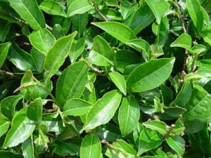
Green tea (Camellia sinensis L. Kuntze) and its main ingredients, epigallocatechin gallate (EGCG, also known as epigallocatechin-3-gallate) and L-theanine, can enhance cognition, neuropsychology and brain functions, as the majority of the reviewed studies suggest [28]. Most of the authors attribute the beneficial effects of green tea to the high levels of EGCG and other antioxidants [29].
The polyphenolic EGCG protects cells from damage associated with oxidative stress and suppresses the activity of pro-inflammatory chemicals produced in your body, such as tumor necrosis factor alpha (TNFα) [30]. This protection can help reduce brain damage that could lead to mental decline and brain diseases such as Parkinson’s and Alzheimer’s. Studies based on functional magnetic resonance imaging (MRI) also found short-term benefits in memory and attention as well as an increase in the activation of a brain area responsible for mediating working memory [29, 30].
The green tea extract used in the MINFIRE formula is of premium quality and uses an advanced extraction process that eliminates the caffeine and retains the antioxidant polyphenols (95% of its total content). Of the total polyphenols in our extract, 80% are phenolic catechins and 50% are pure EGCG. To prepare our green tea extract, a whopping 100 kilograms of raw green tea leaves needs to be used to produce just a single kilogram of the extract.
Choline L-Bitartrate
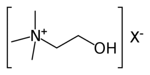
Choline it is neither a vitamin nor a mineral, but it is often grouped with the vitamin B complex due to its similarities. In fact, this nutrient affects a number of vital bodily functions like liver function, muscle movement, metabolism, healthy brain development and nervous system function [31].
As mentioned above, the cholinergic system is modulated by the neurotransmitter called acetylcholine. The synthesis of acetylcholine largely depends on dietary choline intake [32]. Although our body is able to produce choline, we need to get it from the diet to avoid a deficiency. Indeed, many people are not meeting the recommended intake for this nutrient [31]. Example of foods that are source of choline are: liver, egg yolks, meat, fish, broccoli and cauliflower. An alternative for overcoming a possible choline deficiency is through supplementation. Supplementing with choline L-bitartrate is an excellent choice, as it has a much higher bioabsorption rate than plain choline when taken orally in capsules or tablets.
Researchers have been investigated extensively the long-term effects of dietary choline availability on brain and memory. Studies in vivo with rats concluded that the brain’s acetylcholine concentrations increase after an enriched choline diet [33] and memorization of food locations improve after rats were administered with choline either prenatally [34-37], around birth [37, 38] or later in life [39, 40]. In humans, studies show that choline supplementation may help the elderly to recover from moderate Alzheimer’s dementia and other memories impairments [41-44].
L-tirosina
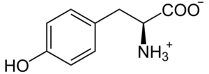
There is a strong relation between L-tyrosine intake and the enhancement of brain catecholamine synthesis in humans [46, 47]. In relation with that, two clinical studies on patients with depression and healthy volunteers have demonstrated that L-tyrosine supplementation positively supports depression management [48]. Moreover, as L-tyrosine increases dopamine levels in the brain, it can improve mental performance in stressful situations. Indeed, one clinical study asked 22 healthy adults to switch between two different tasks rapidly. Compared to the control group, L-tyrosine positively promoted cognitive flexibility (a brain function assumed to be regulated by dopamine) [49].
Other important function of tyrosine is the production of thyroid hormones thyroxine (T4) and the triiodothyronine (T3). T3 and T4 regulate the body’s metabolism of proteins, fats, and carbohydrates, directing how the body uses these compounds to produce energy. The thyroid gland cannot produce these two hormones without the presence of the amino acid L-tyrosine and the mineral iodine. Thus, a deficiency of either the amino acid or the mineral or both can contribute to low thyroid hormone levels. Studies have found L-tyrosine can be beneficial for treating fatigue, a common symptom of low thyroid or adrenal hormone levels [50].
Tyrosine also helps produce melanin, the pigment responsible for hair and skin color [51]. It helps in the function of organs responsible for making and regulating hormones, including the adrenal and pituitary glands. It is involved in the structure of almost every protein in the body.
Phosphatidylserine
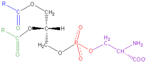
Phosphatidylserine includes two fatty acids that can vary from saturated or monounsaturated to polyunsaturated omega-6 and omega-3 versions like docosahexaenoic acid (DHA) [53]. The DHA is the major polyunsaturated fatty acid in the central nervous system and accumulates particularly in phosphatidylserine. A correct supply of DHA is essential for the synthesis of phosphatidylserine in the body [55].
The concentration of phosphatidylserine in our brain tissues decreases as the years go by and this decrease is related to the deficit of mental capacity and memory [53]. A randomized, double-blind, placebo-controlled clinical trial study concluded that phosphatidylserine supplementation produce significant improvements in Attention-deficit hyperactivity disorder (ADHD), short-term auditory memory and working memory and mental performance to visual stimuli [56]. The effect of the combination of phosphatidylserine and DHA was evaluated in another study in vivo. Such study concluded that this combination can reduced the concentration of damaging reactive oxygen species in brain and liver and may improve learning and memory [57].
The fist scientific studies evaluating the beneficial effect of phosphatidylserine treatment in patients with dementia, were published in the in the early 90’s and were employing bovine brain-derived phosphatidylserine [58]. However, due to concerns regarding bovine spongiform encephalopathy, the most recent studies have based their findings using phosphatidylserine from a vegetal source, mainly obtained by the enzymatic conversion of soybean lecithin (according to the European regulation, only the non-bovine brain-derived phosphatidylserine is allowed in food supplements). Because phosphatidylserine is found in soy lecithin at about 3% of total phospholipids, our ingredient is treated enzymatically in a process called transphosphatidylation to increases its content in phosphatidylserine to 20%.
DHA is one of the components of another amazing Nutribiolite’s product, the OMEGA 3 + VIT K2 + VIT D3. This food supplement has 1000 mg of pure OMEGA 3 fatty acid standardized to 50% of EPA (500 mg) and 25% in DHA (250 mg).
Take care of your brain
-
19.05 €Original price was: 19.05 €.15.36 €Current price is: 15.36 €.
- Stough, C., et al., The chronic effects of an extract of Bacopa monniera (Brahmi) on cognitive function in healthy human subjects. Psychopharmacology (Berl), 2001. 156(4): p. 481-4.
- Kongkeaw, C., et al., Meta-analysis of randomized controlled trials on cognitive effects of Bacopa monnieri extract. J Ethnopharmacol, 2014. 151(1): p. 528-35.
- Pase, M.P., et al., The cognitive-enhancing effects of Bacopa monnieri: a systematic review of randomized, controlled human clinical trials. J Altern Complement Med, 2012. 18(7): p. 647-52.
- Stough, C., et al., Examining the cognitive effects of a special extract of Bacopa monniera (CDRI08: Keenmnd): a review of ten years of research at Swinburne University. J Pharm Pharm Sci, 2013. 16(2): p. 254-8.
- Rastogi, S., R. Pal, and KulshreshthaDk, Bacoside A3-a triterpenoid saponin from Bacopa monniera. Phytochemistry, 1994. 36(1): p. 133-7.
- Bhattacharya, S.K., A. Kumar, and S. Ghosal, Effect of Bacopa monniera on animal models of Alzheimer’s disease and perturbed central cholinergic markers of cognition in rats., in Molecular Aspects of Asian Medicines., D.V. Siva Sankar, Editor. 1999, PJD Publications: New York.
- Bhattacharya, S.K., et al., Antioxidant activity of Bacopa monniera in rat frontal cortex, striatum and hippocampus. Phytother Res, 2000. 14(3): p. 174-9.
- Russo, A., et al., Free radical scavenging capacity and protective effect of Bacopa monniera L. on DNA damage. Phytother Res, 2003. 17(8): p. 870-5.
- Russo, A., et al., Nitric oxide-related toxicity in cultured astrocytes: effect of Bacopa monniera. Life Sci, 2003. 73(12): p. 1517-26.
- Arivazhagan, P., et al., Effect of DL-alpha-lipoic acid on the status of lipid peroxidation and antioxidant enzymes in various brain regions of aged rats. Exp Gerontol, 2002. 37(6): p. 803-11.
- Jackson, C.E., Cholinergic System, in Encyclopedia of Clinical Neuropsychology, J.S. Kreutzer, J. DeLuca, and B. Caplan, Editors. 2011, Springer New York: New York, NY. p. 562-564.
- Nemetchek, M.D., et al., The Ayurvedic plant Bacopa monnieri inhibits inflammatory pathways in the brain. J Ethnopharmacol, 2017. 197: p. 92-100.
- Viji, V. and A. Helen, Inhibition of pro-inflammatory mediators: role of Bacopa monniera (L.) Wettst. Inflammopharmacology, 2011. 19(5): p. 283-91.
- Thongbai, B., et al., Hericium erinaceus, an amazing medicinal mushroom. Mycological Progress, 2015. 14(10): p. 91.
- Lu, Q.-Q., et al., Bioactive metabolites from the mycelia of the basidiomycete Hericium erinaceum. Natural Product Research, 2014. 28(16): p. 1288-1292.
- Zhang, C.C., et al., Chemical constituents from Hericium erinaceus and their ability to stimulate NGF-mediated neurite outgrowth on PC12 cells. Bioorg Med Chem Lett, 2015. 25(22): p. 5078-82.
- Wang, K., et al., Erinacerins C–L, Isoindolin-1-ones with α-Glucosidase Inhibitory Activity from Cultures of the Medicinal Mushroom Hericium erinaceus. Journal of Natural Products, 2015. 78(1): p. 146-154.
- Li, J.L., et al., A comparative study on sterols of ethanol extract and water extract from Hericium erinaceus. Zhongguo Zhong Yao Za Zhi, 2001. 26(12): p. 831-4.
- Friedman, M., Chemistry, Nutrition, and Health-Promoting Properties of Hericium erinaceus (Lion’s Mane) Mushroom Fruiting Bodies and Mycelia and Their Bioactive Compounds. J Agric Food Chem, 2015. 63(32): p. 7108-23.
- Aloe, L., et al., Nerve growth factor: from the early discoveries to the potential clinical use. Journal of Translational Medicine, 2012. 10(1): p. 239.
- DeFeudis, F.V. and K. Drieu, Ginkgo Biloba Extract (EGb 761) and CNS Functions Basic Studies and Clinical Applications. Current Drug Targets, 2000. 1(1): p. 25-58.
- Diamond, B.J., et al., Ginkgo biloba extract: mechanisms and clinical indications. Arch Phys Med Rehabil, 2000. 81(5): p. 668-78.
- Kubota, Y., et al., Effects of Ginkgo biloba extract on blood pressure and vascular endothelial response by acetylcholine in spontaneously hypertensive rats. Journal of Pharmacy and Pharmacology, 2006. 58(2): p. 243-249.
- Spinnewyn, B., et al., Involvement of platelet-activating factor (PAF) in cerebral post-ischemic phase in Mongolian gerbils. Prostaglandins, 1987. 34(3): p. 337-349.
- McKenna, D.J., K. Jones, and K. Hughes, Efficacy, safety, and use of ginkgo biloba in clinical and preclinical applications. Altern Ther Health Med, 2001. 7(5): p. 70-86, 88-90.
- Ernst, E., The Risk–Benefit Profile of Commonly Used Herbal Therapies: Ginkgo, St. John’s Wort, Ginseng, Echinacea, Saw Palmetto, and Kava. Annals of Internal Medicine, 2002. 136(1): p. 42-53.
- Nathan, P., Can the cognitive enhancing effects of Ginkgo biloba be explained by its pharmacology? Medical Hypotheses, 2000. 55(6): p. 491-493.
- Mancini, E., et al., Green tea effects on cognition, mood and human brain function: A systematic review. Phytomedicine, 2017. 34: p. 26-37.
- Higdon, J.V. and B. Frei, Tea Catechins and Polyphenols: Health Effects, Metabolism, and Antioxidant Functions. Critical Reviews in Food Science and Nutrition, 2003. 43(1): p. 89-143.
- Scapagnini, G., et al., Modulation of Nrf2/ARE Pathway by Food Polyphenols: A Nutritional Neuroprotective Strategy for Cognitive and Neurodegenerative Disorders. Molecular Neurobiology, 2011. 44(2): p. 192-201.
- Zeisel, S.H. and K.-A.d. Costa, Choline: an essential nutrient for public health. Nutrition reviews, 2009. 67(11): p. 615-623.
- Lippelt, D.P., et al., No Acute Effects of Choline Bitartrate Food Supplements on Memory in Healthy, Young, Human Adults. PloS one, 2016. 11(6): p. e0157714-e0157714.
- Hirsch, M.J. and R.J. Wurtman, Lecithin Consumption Increases Acetylcholine Concentrations in Rat Brain and Adrenal Gland. Science, 1978. 202(4364): p. 223-225.
- Moon, J., et al., Perinatal choline supplementation improves cognitive functioning and emotion regulation in the Ts65Dn mouse model of Down syndrome. Behav Neurosci, 2010. 124(3): p. 346-61.
- Velazquez, R., et al., Maternal choline supplementation improves spatial learning and adult hippocampal neurogenesis in the Ts65Dn mouse model of Down syndrome. Neurobiology of Disease, 2013. 58: p. 92-101.
- Meck, W.H. and C.L. Williams, Perinatal choline supplementation increases the threshold for chunking in spatial memory. Neuroreport, 1997. 8(14): p. 3053-9.
- Meck, W.H., R.A. Smith, and C.L. Williams, Organizational changes in cholinergic activity and enhanced visuospatial memory as a function of choline administered prenatally or postnatally or both. Behavioral Neuroscience, 1989. 103(6): p. 1234-1241.
- Schenk, F. and C. Brandner, Indirect effects of peri- and postnatal choline treatment on place-learning abilities in rat. Psychobiology, 1995. 23(4): p. 302-313.
- Mizumori, S.J., et al., Effects of dietary choline on memory and brain chemistry in aged mice. Neurobiol Aging, 1985. 6(1): p. 51-6.
- Tees, R.C., The influences of sex, rearing environment, and neonatal choline dietary supplementation on spatial and nonspatial learning and memory in adult rats. Dev Psychobiol, 1999. 35(4): p. 328-42.
- Scapicchio, P.L., Revisiting choline alphoscerate profile: a new, perspective, role in dementia? Int J Neurosci, 2013. 123(7): p. 444-9.
- Traini, E., V. Bramanti, and F. Amenta, Choline alphoscerate (alpha-glyceryl-phosphoryl-choline) an old choline- containing phospholipid with a still interesting profile as cognition enhancing agent. Curr Alzheimer Res, 2013. 10(10): p. 1070-9.
- De Jesus Moreno Moreno, M., Cognitive improvement in mild to moderate Alzheimer’s dementia after treatment with the acetylcholine precursor choline alfoscerate: a multicenter, double-blind, randomized, placebo-controlled trial. Clin Ther, 2003. 25(1): p. 178-93.
- Parnetti, L., F. Amenta, and V. Gallai, Choline alphoscerate in cognitive decline and in acute cerebrovascular disease: an analysis of published clinical data. Mech Ageing Dev, 2001. 122(16): p. 2041-55.
- Blanco, A. and G. Blanco, Chapter 16 – Amino Acid Metabolism, in Medical Biochemistry, A. Blanco and G. Blanco, Editors. 2017, Academic Press. p. 367-399.
- Agharanya, J.C., R. Alonso, and R.J. Wurtman, Changes in catecholamine excretion after short-term tyrosine ingestion in normally fed human subjects. Am J Clin Nutr, 1981. 34(1): p. 82-7.
- Fernstrom, J. and M. Fernstrom, Tyrosine, phenylalanine, and catecholamine synthesis and function in the brain. The Journal of nutrition, 2007. 137 6 Suppl 1: p. 1539S-1547S; discussion 1548S.
- Alabsi, A., A.C. Khoudary, and W. Abdelwahed, The Antidepressant Effect of L-Tyrosine-Loaded Nanoparticles: Behavioral Aspects. Annals of neurosciences, 2016. 23(2): p. 89-99.
- Steenbergen, L., et al., Tyrosine promotes cognitive flexibility: evidence from proactive vs. reactive control during task switching performance. Neuropsychologia, 2015. 69: p. 50-5.
- Watson, P., Tyrosine supplementation: Can this amino acid boost brain dopamine and improve physical and mental performance? Sports Science Exchange, 2016. 28(157): p. 1-6.
- Rzepka, Z., et al., From tyrosine to melanin: Signaling pathways and factors regulating melanogenesis. Postepy Hig Med Dosw (Online), 2016. 70(0): p. 695-708.
- Glade, M.J. and K. Smith, Phosphatidylserine and the human brain. Nutrition, 2015. 31(6): p. 781-786.
- Kim, H.-Y., B.X. Huang, and A.A. Spector, Phosphatidylserine in the brain: metabolism and function. Progress in lipid research, 2014. 56: p. 1-18.
- Wheeler, K.P. and R. Whittam, The involvement of phosphatidylserine in adenosine triphosphatase activity of the sodium pump. J Physiol, 1970. 207(2): p. 303-28.
- Garcia, M.C., et al., Effect of Docosahexaenoic Acid on the Synthesis of Phosphatidylserine in Rat Brain Microsomes and C6 Glioma Cells. Journal of Neurochemistry, 1998. 70(1): p. 24-30.
- Hirayama, S., et al., The effect of phosphatidylserine administration on memory and symptoms of attention-deficit hyperactivity disorder: a randomised, double-blind, placebo-controlled clinical trial. Journal of Human Nutrition and Dietetics, 2014. 27(s2): p. 284-291.
- Chaung, H.-C., et al., Docosahexaenoic acid and phosphatidylserine improves the antioxidant activities in vitro and in vivo and cognitive functions of the developing brain. Food Chemistry, 2013. 138(1): p. 342-347.
- Amaducci, L., et al., Use of phosphatidylserine in Alzheimer’s disease. Ann N Y Acad Sci, 1991. 640: p. 245-9.
Subscribe to our newsletter to save 5% on your first order.
Thank you!
You have successfully joined us. Please check your email with the discount code.
